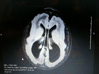
Ok. I'm ready. I'm about to post pictures of Keira's MRI. These pictures were taken when she was about 4 weeks old. If you are interested, I would like you to see the damage done by CMV to her brain. I am not going to use technical terms. Partly, you don't need it to see the difference, partly because I'd have to go back to my textbooks to figure it out.
Here is an MRI of a normal brain. The comparison pictures are an adult, but for my purposes here, it's a fair comparison. Sorry I'm not tech-saavy enough to make my own little pointers. Bare with me.
This brain is nice and compact. There is a little fluid/space (the black part) between the brain and the skull, but not much. There are lots of squiggles (that's where most of the info is stored). See the sideways "C" in the middle? See the little bit of black inside the C in the very center? That, I understand, is the ventricles. It's fluid, and there is not a lot of it.
Here is Keira's MRI:
What do you see? There is more black, or fluid between her scull and her brain, particularly toward the back (parietal and occipital lobes). There are very few squiggles. The ventricles are HUGE.
I feel a little sick to my stomach just looking at it again.
Of course we hope and pray that with therapy, therapy, therapy, her brain is rejuvenating itself somewhat. Hopefully there are a couple more wrinkles and a little less fluid. We may never know unless we need an MRI again for some other reason. At this point another MRI would be for curiosity's sake only...what she can do is what she can do, regardless of what the pictures look like.
More than one therapist and/or doctor have commented, when meeting Keira after first reviewing her records, that she is doing much better than they may have expected. We'll take what we can get.
Two more, just to compare:


No comments:
Post a Comment